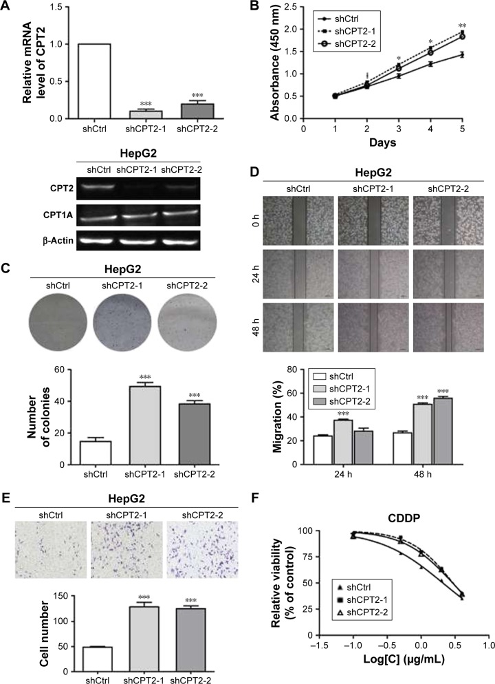
Figure 2.
CPT2-depleted HepG2 cells displayed increased cell proliferation, metastasis capabilities, and chemoresistance to cisplatin. (A) CPT2 expression was confirmed by qRT-PCR and Western blot. (B) CCK-8 assay and (C) colony formation assay indicate that growth rates are increased in CPT2-repressing HepG2 cells. (D) wound-healing assay and (E) transwell assay with matrigel showing that cell invasion capability was remarkably increased in CPT2-repressing cells compared with the control group. (F) CPT2 silencing rendered HepG2 cells more resistant to cisplatin. ShCtrl and shCPT2-1/2 stably knocked down HepG2 cells were treated with different concentrations of cisplatin (0.1, 0.5, 1.0, 2.0, and 4.0 μg/mL) for 72 h, and then the cell viability was detected by MTT. Data are presented as mean ± SD of three independent experiments. *P<0.05; **P<0.01; ***P<0.001.
Abbreviations: CPT2, carnitine palmitoyltransferase-2; qRT-PCR, quantitative reverse transcriptase polymerase chain reaction; CCK-8, Cell Counting Kit-8; MTT, 3-(4,5-dimethyl-2-thiazolyl)-2,5-diphenyl-2H-tetrazolium bromide.

