Fig. 3.
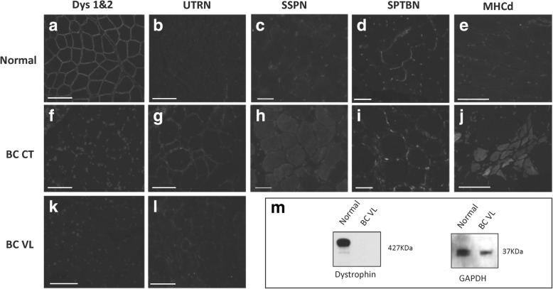
Dystrophin deficiency in the affected border collie (BC) dog. Normal and dystrophic muscle were immunostained for DYS1 and 2 (a, f, k). Peri-membranous dystrophin expression was seen in each myofiber of normal muscle (a) but was absent in the affected dog (f, k). Utrophin (UTRN) was minimally expressed in normal muscle (b) but, by comparison, was increased in the affected dog (g, l). Similarly, sarcospan (SSPN) was minimally expressed in normal muscle (c) and comparably increased in the affected dog (h). Spectrin (SPTBN) was used as a cellular membrane marker (d, i). Myosin heavy chain developmental fibers (MHCd) positive myofibers were absent in normal muscle (e) but present in the affected dog (j). Nuclei were stained with DAPI. All images were taken with a × 20 objective. m Western blot showed absent dystrophin in the BC; GAPDH was used as a loading control. Metric bar = 100 μm

