
Fig. 1.
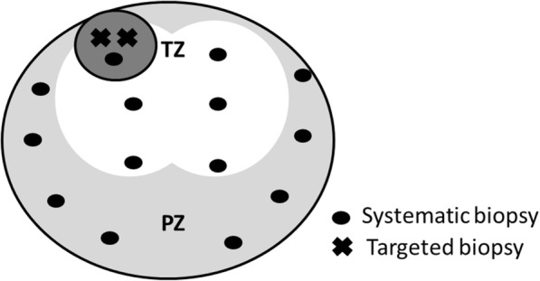
Biopsy strategy. In the MRI cohort, 12 to 14 cores were biopsied. In patients who had suspicious lesions on mpMRI, each suspicious lesion could be targeted as one of systematic biopsy at the nearest point and further typically two targeted biopsies were added for each lesion. White, light gray, and dark gray areas indicate transitional zone, peripheral zone of axial view, and index lesion on mpMRI, respectively. Black dot indicates systematic biopsy cores and x indicates targeted biopsy cores. TZ; transitional zone, PZ: peripheral zone