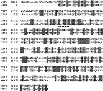
Figure 3.
Comparison of C. neoformans Uxs1 to S. typhimurium Orf3. Identical amino acids are shaded dark gray and similar residues are shaded light gray. The NAD+ binding motif of Uxs1 is underscored with * and is aligned with a similar motif in the bacterial sequence that falls in a well-conserved region. The N-terminal 200 residues of Orf3 are not shown.

