Figure 2.
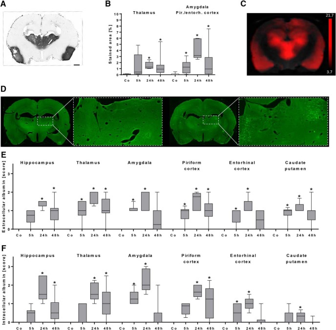
Histologic evaluation of albumin extravasation during early epileptogenesis. A, Distribution of FITC-labeled albumin (FITC-Alb) after its conversion into a light microscopically visible adduct with anti-FITC-HRP and nickel-enhanced DAB in a representative section from a rat 48 h following status epilepticus (SE). The extravasation marker indicating leakage of allocated BBB is predominantly visible in the thalamus and the piriform cortex. Scale bar = 1 mm. B, Quantification of DAB-positive area relative to the total section area in control rats (n = 10) and rats 5 h (n = 6), 24 h (n = 5), and 48 h (n = 16) following SE. * p < 0.05 compared to control, one-way ANOVA, Dunnett’s multiple comparison test. C, Coronal Gd-DTPA-enhanced T1 MRI leakage map resulting from comparison between baseline and 48 h post-SE. Note the striking similarity of BBB leakage pattern in the ex vivo DAB-converted FITC-albumin slice (A) and in vivo contrast-enhanced MRI (C). Leakage t-map was calculated by SPM12 software (two-sample unpaired t test, p < 0.001, and a minimum cluster size of 100 voxels; scale bar displays t-values). D, Exemplary whole-brain sections and a higher-magnified image of the thalamus from two rats, predominantly showing extracellular distribution (left, 48 h post-SE) or intracellular uptake (right, 24 h post-SE) of extravasated FITC-labeled albumin. E, Semiquantitative analysis of extracellular FITC-labeled albumin in control rats (n = 10) and rats 5 h (n = 6), 24 h (n = 5), and 48 h (n = 14) following SE. F, Semiquantitative analysis of intracellular FITC-labeled albumin in control rats (n = 10) and rats 5 h (n = 6), 24 h (n = 5), and 48 h (n = 14) following SE. E and F show peak values of FITC-albumin presence at 24 h post-SE. * p < 0.05 compared to control, Kruskal–Wallis ANOVA, Dunn’s multiple comparison test. Data are illustrated as box-and-whisker plots. Co, control, Pir./entorh., piriform/entorhinal.

