
Figure 4.
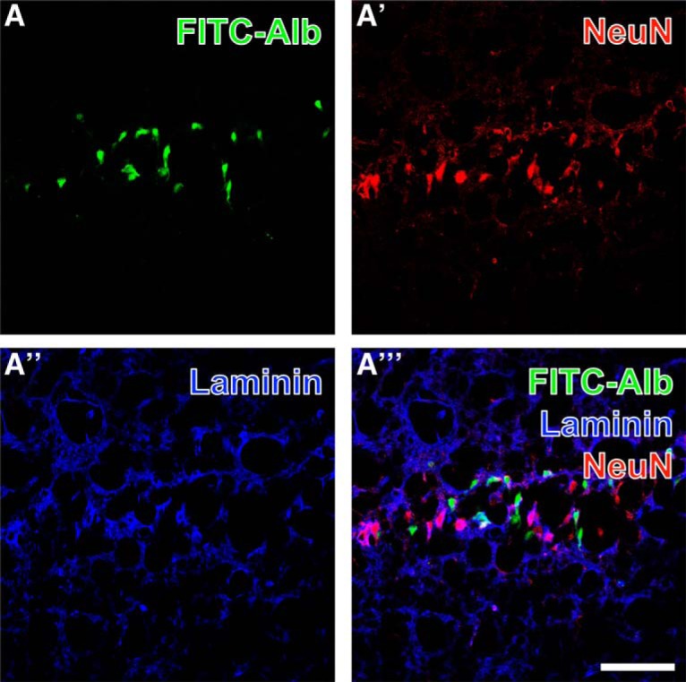
Concomitant detection of FITC-coupled albumin (FITC-Alb; A) in the pyramidal layer of the hippocampal CA1 region 48 h after SE shows predominantly intracellular FITC-Alb. Neuronal somata are stained with biotinylated anti-NeuN and red fluorescent Cy3-streptavidin (A′), while vascular basement membranes are visualized with rabbit anti-laminin and Cy5-donkey anti-rabbit IgG (A″, immunosignals color-coded in blue). The overlay of staining patterns (A″′) elucidates only rare colocalization of FITC-Alb and NeuN-positive neurons. Scale bar = 50 µm.