
Figure 5.
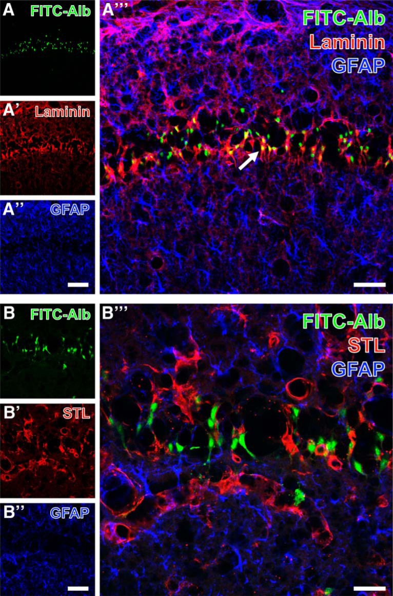
Hippocampal pyramidal cell layer in the CA1 region with FITC-coupled albumin (FITC-Alb; A, B) 48 h following SE and laminin-immunoreactivity in vascular basement membranes (Cy3, red; A′) or endothelial binding sites for biotinylated Solanum tuberosum lectin (STL; Cy3, red; B′), each combined with immunolabeling of astroglial GFAP (A″, B″, Cy5, color-coded in blue) at lower (A–A″′) and higher (B–B″′) magnification. Merged staining patterns (A″′, B″′) elucidate FITC-Alb in close vicinity to laminin-immunopositive structures (arrow in A″′), but no obvious colocalization of FITC-Alb and GFAP-positive astrocytes (A″′, B″′). Scale bars: A″ (also valid for A, A′) = 100 µm, A″′ = 50 µm, B″ (also valid for B, B′) = 50 µm, A″′ = 25 µm.