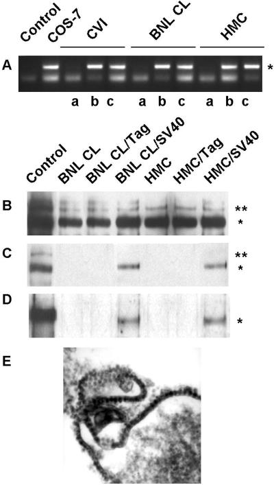
Figure 4.
Coculture experiments. (A) Tag sequence (*) was amplified by PCR from genomic DNA of CV-1, BNL CL, and HMC. PCR without DNA (lane 1) and with COS-7 DNA (lane 2) were also performed as controls. Shown is the product of PCR amplification performed on lysates from cells cocultured with Tag-HMC (a) and SV40-HMC (b). The same PCR amplification was also performed on the SV40-HMC coculture medium (c). (B) Solubilized proteins from cell lysates were immunoprecipitated with Met antibodies and probed with the same Met antibodies and (C) with the antiphosphotyrosine antibodies. Asterisks on the right indicate the positions of the pr170MET precursor (**) and p145MET (*). (D) Tag protein (*) was detected in cell lysates immunoprecipitated and probed with Tag antibodies. (E) Electron microscopy of SV40-HMC cells. (Original magnification: ×60,000.)

