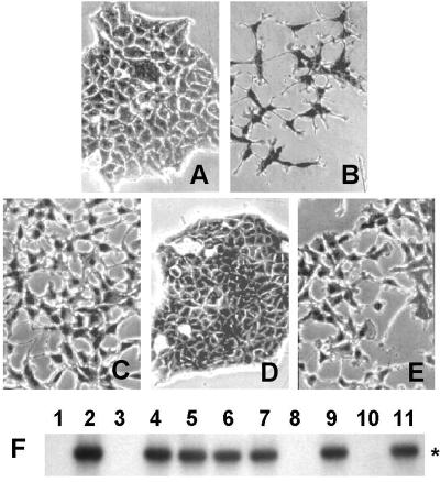
Figure 6.
Met/HGF autocrine loop. Scatter assay was performed on the MDCK cell line. Shown are untreated MDCK cells (A) and MDCK stimulated with recombinant HGF (B), with MMP-conditioned medium (C), with MMM-conditioned medium (D), and with spBNL-conditioned medium (E). (Original magnification: ×320.) (F) RT-PCR and Southern hybridization specific for HGF (nucleotides 646-1533). Cell lines expressing activated Met (MMP, lane 4; MMCa, lane 5; Tag-HMC, lane 6; SV40-HMC, lane 7; HMC/SV40, lane 9; and spBNL, lane 11) display HGF expression (*), whereas HGF cDNA was not amplified in MMM (lane 3), HMC/Tag (lane 8), and BNL CL (lane 10) cells as well as in RT-PCR control without RNA (lane 1). MRC5 cell line (lane 2) was used as a control of HGF expression.

