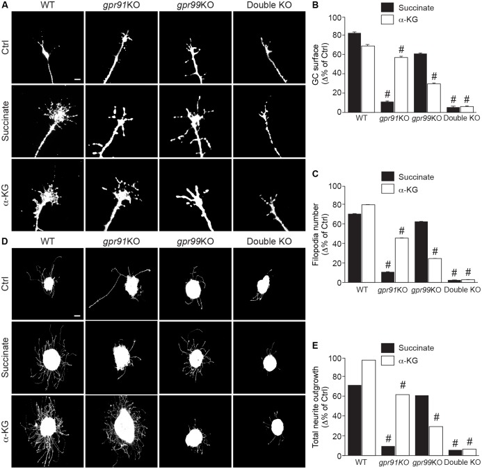
Fig 2. Succinate and α-KG modulate GC morphology and increase neurite outgrowth via GPR91 and GPR99.
(A) Photomicrographs of E14/15 GCs of retinal projections from WT, gpr91KO, gpr99KO, and double-KO mouse embryos, untreated and after treatment with GPR91 agonist (100 μM succinate) or GPR99 agonist (200 μM α-KG). (B) Analysis of GC surface area (N = 104–271 per condition) and (C) filopodia number (N = 105–271 per condition) following a 1 h treatment with GPR91 or GPR99 ligands in WT, gpr91KO, gpr99KO, and double-KO mice. (D) Photomicrographs of retinal explants from each mouse strain cultured for 1 DIV and treated for 15 h with succinate (100 μM) or α-KG (200 μM). (E) Quantification of neurite growth after treatment with GPR91 or GPR99 agonist (N = 84–181 per condition). Scale bars: 5 μm (A); 100 μm (D). Values are presented as the means ± SEM. # indicates significant changes compared to WT in B, C, and E; p < 0.001. Underlying data can be found in S1 Data. α-KG, α-ketoglutarate; Ctrl, control; DIV, day in vitro; E14/15, embryonic day 14/15; GC, growth cone; KO, knockout; WT, wild-type.

