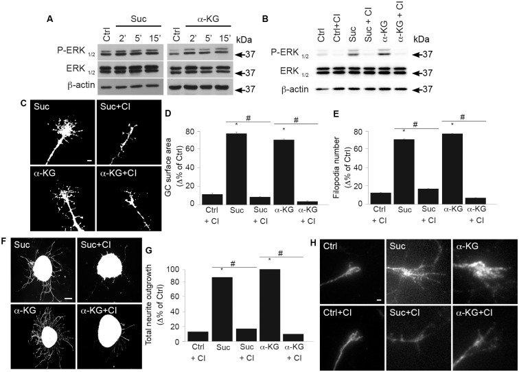
Fig 3. GPR91 and GPR99 agonists modulate GC morphology and increase axon growth via the ERK1/2 pathway.
(A) Protein expression levels of P-ERK1/2, ERK1/2, and β-actin in primary neuron cultures incubated with one of the following: 100 μM succinate or 200 μM α-KG at 37 °C for 2, 5, and 15 min. The antibody β-actin was used to verify equal loading in all lanes. (B) ERK phosphorylation state following 15 min pretreatment with CI-1040, an ERK1/2 inhibitor, before incubation with or without GPR91 or GPR99 agonist. (C) Photomicrographs of retinal explant GCs treated with GPR91 or GPR99 agonist in the presence or absence of CI-1040. (D) Quantification of the GC surface areas (N = 178–189 per condition) and (E) filopodia numbers (N = 178–237 per condition). (F) Photomicrographs of retinal explants treated with succinate (100 μM) or α-KG (200 μM) in the presence or absence of CI-1040. (G) Analysis of retinal projection growth of retinal explants (N = 31–86 per condition). (H) Immunohistochemical photomicrographs of the ERK1/2 phosphorylation state in retinal explant GCs treated with 100 μM succinate or 200 μM α-KG in the presence or absence of CI-1040. Scale bars: 5 μm (C, H); 100 μm (F). Values are presented as the means ± SEM. * indicates a significant change compared to the Ctrl group in D, E and G; p < 0.0001. # indicates a significant change induced by ERK inhibitor treatment in D, E and G; p < 0.002. Underlying data can be found in S1 Data. α-KG, α-ketoglutarate; Ctrl, control; ERK1/2, extracellular signal–regulated kinases 1 and 2; GC, growth cone.

