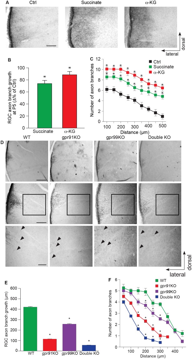
Fig 4. Succinate/GPR91 and α-KG/GPR99 are involved in RGC axon growth in vivo.
(A) Photomicrographs of P5 hamster retinal projections in the DTN for the control, succinate (100 mM), and α-KG (200 mM) treatment groups. (B, C) Quantification of retinal projection development in the DTN: collateral axon projection length (N = 168–240 per condition) and collateral axon branch number (N = 34–57 per condition) of treated groups compared with the Ctrl group. Succinate and α-KG treatment increased axon growth and collateral branch number compared with the Ctrl. (D) Photomicrographs of P5 retinal projections in the DTN in WT, gpr91KO, gpr99KO, and double-KO mice. (E) Quantification of retinal projections in the DTN during the development of WT, gpr91KO, gpr99KO, and double-KO mice (N = 627–963 per condition). Collateral projection length is expressed as the mean ± SEM. (F) The number of collateral axon branches decreased in gpr91KO, gpr99KO, and double-KO mice compared to WT (N = 82–160 per condition). Data are presented as the means ± SEM. Scale bars: 100 μm (A, D). * indicates a significant change compared to the Ctrl group in B and C (p < 0.05) and in E and F (p < 0.0001). Underlying data can be found in S1 Data. α-KG, α-ketoglutarate; Ctrl, control; DTN, dorsal terminal nucleus; KO, knockout; RGC, retinal ganglion cell; WT, wild-type.

