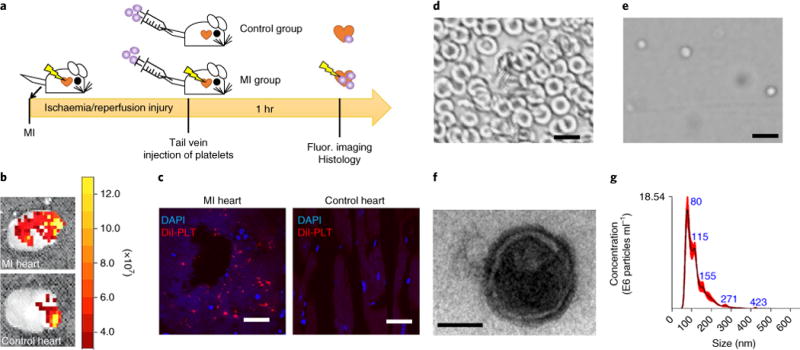
Fig. 1. Platelet binding to myocardial infarction sites and the derivation of platelet nanovesicles.

a, A schematic showing the animal study design to test the innate binding ability of platelets to sites of myocardial infarction (MI). b, Representative ex vivo fluorescent imaging showing binding of intravenously injected DiI-labelled platelets in hearts with or without ischaemia/reperfusion (I/R) injury. c, Representative fluorescent microscopic images showing the targeting of Dil-labelled platelets (red) to the MI area (DAPI, nuclei). Scale bars, 100 μm. d,e, Collected rat red blood cells (d) as seen under a light microscope, demonstrating a distinctive morphology compared to platelets (e). Scale bars, 10 μm. f, A transmission electron micrograph of a platelet nanovesicle. Scale bar, 100 nm. g, Size examination of platelet membrane nanovesicles by NanoSight.
