
Fig. 2. Generation and characterization of PNV-CSCs.
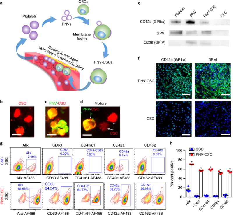
a, A schematic showing the overview of PNV decoration and PNV-CSC therapy. b,c, Red fluorescent DiI-labelled CSCs (b) were fused with green fluorescent DiO-labelled PNVs to form PNV-CSCs (c). d, Co-incubation of CSCs (red) with PNV-CSCs (yellow). Scale bar, 20 μm. e, Western blot analysis revealed the expressions of platelet-specific markers including CD42b (GPIbα), GPVI and CD36 (GPIV) in platelets, PNVs and PNV-CSCs, but not in CSCs. Original western blot images can be found in Supplementary Figs. 2–4. f, Immunocytochemistry staining confirmed CD42b (GPIbα) and GPVI expression in PNV-CSCs (top), but not in CSCs (bottom). Scale bars, 200 μm. g,h, Flow cytometric analysis of platelet and exosome surface marker expressions on PNV-CSCs (n = 3) and CSCs (n = 4). *P< 0.05. All values are mean ± s.d. Two-tailed t-test for comparison.