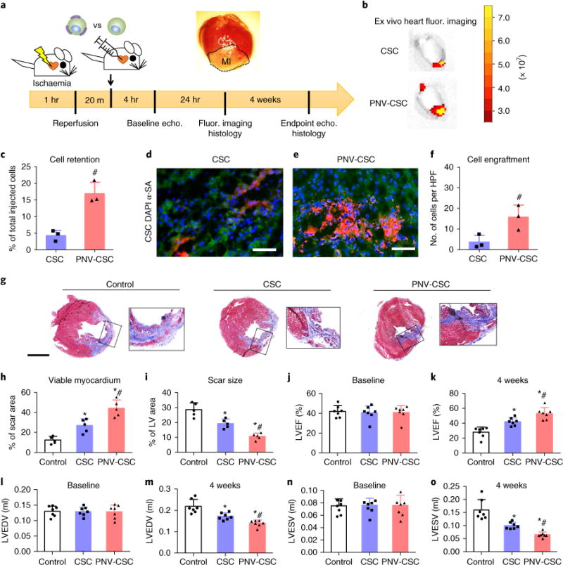
Fig. 5. PNV decoration boosts CSC retention and therapeutic outcomes in rats with myocardial infarction.

a, A schematic showing the animal study design. b, Representative ex vivo fluorescent imaging of ischaemia/reperfusion rat hearts 24 hrs after intracoronary infusion of DiI-labelled PNV-CSCs or CSCs. c, qPCR analysis revealed higher retention rates of PNV-CSCs as compared to those of CSCs (n = 3 animals/hearts per group). d,e, Representative fluorescent micrographs showing engrafted CSCs (d) or PNV-CSCs (e) in the post-MI hearts (green, α-SA antibody). Scale bars, 50 μm. f, Quantitative analysis of cell engraftment by histology (n = 3 animals/hearts per group). HPF, high-power field. g, Representative Masson’s trichrome-stained myocardial sections 4 weeks after treatment (blue, scar tissue; red, viable myocardium). Scale bar, 2 mm. h,i, Quantitative analyses of viable myocardium and scar size from the Masson’s trichrome images (n = 5 animals per group). LV, left ventricle. j,k, Left ventricular ejection fractions (LVEFs) measured by echocardiography at baseline (4 hrs post-MI) and 4 weeks later (n = 7 animals per group). l–o, Measurement of LV end-systolic (LVESV) and end-diastolic (LVEDV) volumes. *P<0.05 when compared to the control group; #P<0.05 when compared to the CSC groups. All values are mean ± s.d. Two-tailed t-test for comparison between two groups. One-way ANOVA with post-hoc Bonferroni test for comparisons that involve three or more groups.
