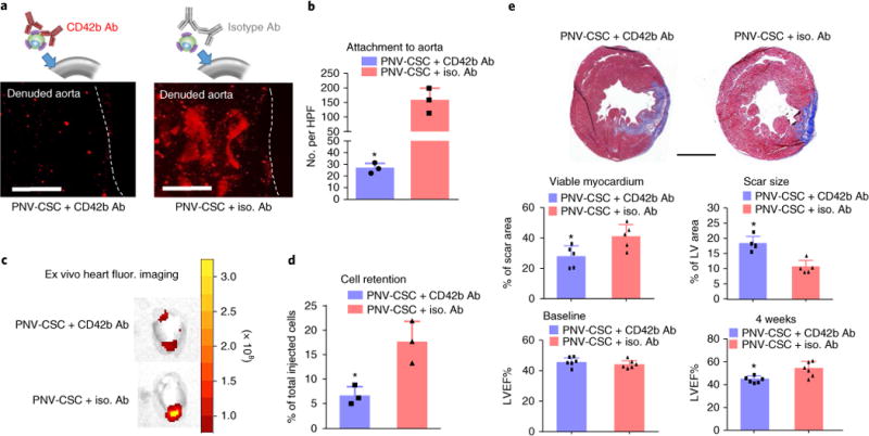
Fig. 7. The role of CD42b in targeting PNV-CSCs to Mi injury.

a, Representative fluorescent micrographs showing the adherence of anti-CD42b or isotype antibody pre-treated PNV-CSCs on denuded rat aortas. b, Quantitation of binding (n = 3 samples per group). HPF, high-power field. c, Representative ex vivo fluorescent imaging of ischaemia/reperfusion rat hearts 24 hrs after intracoronary infusion of anti-CD42b or isotype antibody pre-treated PNV-CSCs. d, Quantitation of cell retention by qPCR (n = 3 animals per group). e, Representative Masson’s trichrome-stained myocardial sections 4 weeks after treatment (blue, scar tissue; red, viable myocardium). Scale bar, 2 mm. Quantitative analyses of viable myocardium and scar size from the Masson’s trichrome images (n = 5 animals per group). Left ventricular ejection fractions (LVEFs) measured by echocardiography at baseline (4 hrs post-MI) and 4 weeks later (n = 6 animals per group). *P< 0.05 when compared to the PNV-CSC + iso. Ab group. All values are mean ± s.d. Two-tailed t-test for comparison.
