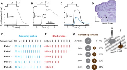
Fig. 1. Design of probe stimuli protocols.

Schematics of eyelid conditioning training (A to C) and probe protocols (D to G). For (A) to (C), schematics of stimuli presentation is shown at the top, and eyelid position as a function of time is shown below. Upward deviation corresponds to eyelid closure. (A) Neutral trained input (CS) is paired with eyelid stimulation as US. In the naïve state, there is no eyelid response during CS, and the US evokes a reflexive noncerebellar eyelid closure (shown in gray). (B) Behavioral response after animal learned CS-US pairing. Predictive eyelid closure during CS and before US is a CR. (C) Example eyelid CR profiles on the CS-alone trial from subject trained to produce either a full-sized (black line) or half-sized eyelid closure (blue line). (D) Coronal histology demonstrating lesion marks from two stimulation electrodes implanted in middle cerebellar peduncle. (E to G) Schematics of probe inputs on three different probe protocols. In each case, input used for training (500-ms, 100-Hz pulse train) is shown in black. (E) Frequency probes. The length of stimulus was kept at 500 ms, but frequency was systematically decreased. (F) Short probes. Frequency was kept at 100 Hz, but only portion of the stimulus length was presented. (G) Competing stimulus. Two separate stimulating electrodes were implanted into middle cerebellar peduncle spaced 1 mm laterally. Only electrode A was used for training. During probe trials, neither frequency nor length of the stimulus was changed, but rather the current applied on electrode A was decreased and, correspondingly, the current on electrode B was increased from zero to keep the number of total activated mossy fibers approximately constant. This manipulation should result in a gradual shift of the overlap between mossy fibers activated by probe and trained stimulus. The area of current spread is illustrated as a gray circle for electrode A (used to deliver trained input) and a brown circle for electrode B (competing stimulus).
