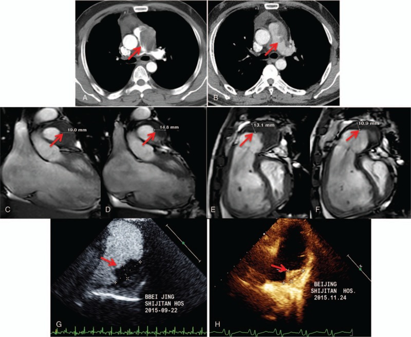
Figure 4.

Imaging changes of before and after treatment. During 31 months follow-up, pulmonary CT demonstrated the lesion of pulmonary artery IgG4-RD in patient 1 shrunk obviously (A: before treatment, B: after treatment). During 3 months follow-up, MRI (C and E: before treatment, D and F: after treatment) and ultrasonic cardiogram (G: before treatment, H: after treatment) showed the lesion of pulmonary artery IgG4-RD also shrunk obviously in patient 3. CT = computed tomography; IgG4-RD = IgG4-related disease; MRI = magnetic resonance imaging.
