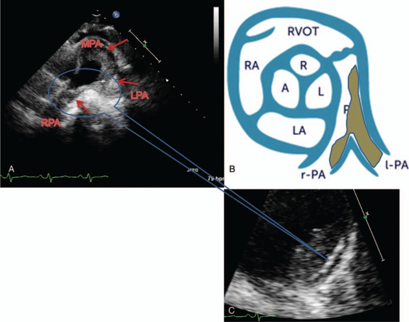
Figure 6.

The presentation of ultrasonic cardiogram in pulmonary artery IgG4-RD causing PH. Ultrasonic cardiogram showed hypoecho lesion of lateral wall in main pulmonary artery (MPA) in patient 3, which extended to the left pulmonary artery (LPA), causing LPA occlusion. Hypoecho lesion extended to the right pulmonary artery (RPA), which induced severe RPA stenosis (A). Enlarge watching, the endothelium of hypoecho lesion was obviously thickening in pulmonary artery, and even destructive (C). The above presentation of ultrasonic cardiogram is like diagram as shown (B). IgG4-RD = IgG4-related disease, PH = pulmonary hypertension.
