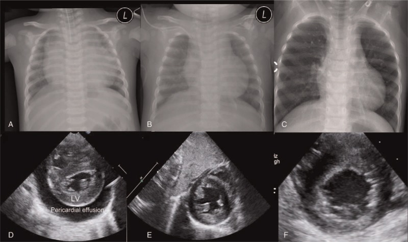
Figure 1.

Patient chest radiographs and echocardiographs. (A–C) Chest x-rays showing severe cardiomegaly (A) that was markedly improved after 2 months (B) and 1 year (C) of dietary intervention. (D–F) A parasternal short axis view of the conducted echocardiograph, showing severe pericardial effusion and left ventricular hypertrophy (D) that was completely resolved after 2 months (E) and 1 year (F) of dietary intervention. L = patient's left side, LV = left ventricle.
