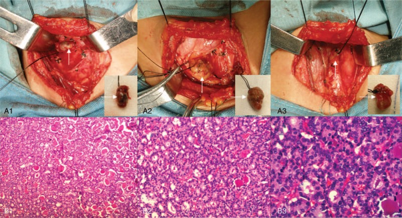
Figure 1.

Parathyroidectomy and pathologic analysis. The first operation involved resection of 3 parathyroid glands (A1–3, arrows) in the normal position. Pathologic analysis showed nodular parathyroid hyperplasia with interstitial fibrosis and calcification (B1–3). Hematoxylin and eosin staining (magnification, ×100 for B1, ×200 for B2, ×400 for B3).
