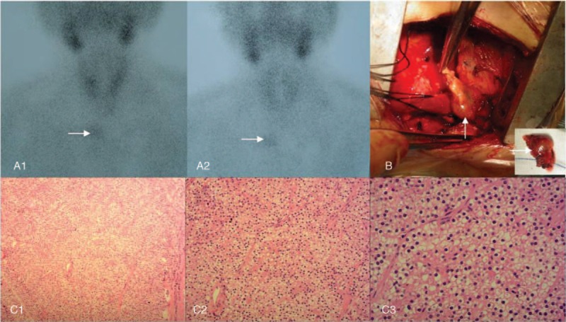
Figure 2.

Ectopic parathyroidectomy and pathologic analysis. Before the second operation, technetium-99m methoxyisobutylisonitrile (99mTc-MIBI) parathyroid scintigraphy showed a single ectopic parathyroid gland in the superior mediastinum (A1–2, arrows). Intraoperatively, the ectopic parathyroid gland in the deep side of the right acromioclavicular joint (B) was removed. Pathologic analysis of the resected specimen showed nodular hyperplasia of parathyroid tissue which was well differentiated (C1–3). Hematoxylin and eosin staining (magnification, ×100 for C1, ×200 for C2, ×400 for C3).
