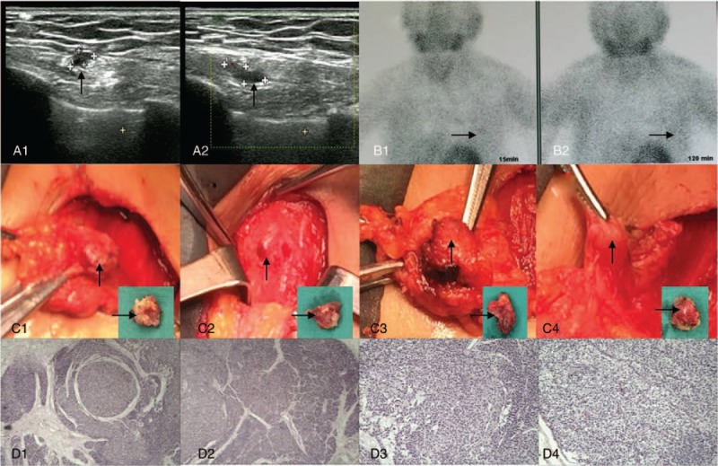
Figure 4.

Partial excision of autotransplanted parathyroid gland and pathologic analysis. Before the fourth operation, B-ultrasound revealed multiple hypoechoic nodules (A1–2, arrows), and technetium-99m methoxyisobutylisonitrile (99mTc-MIBI) parathyroid scintigraphy showed increased uptake of soft tissue lesions (B1–2, arrows). Intraoperatively, 4 markedly enlarged autotransplanted parathyroid glands (C1–4, arrows) were resected. Pathologic analysis showed secondary hyperplasia of the parathyroid (D1–4) in striated muscle tissue. Hematoxylin and eosin staining (magnification, ×40 for D1 and D2, ×100 for D3 and D4).
