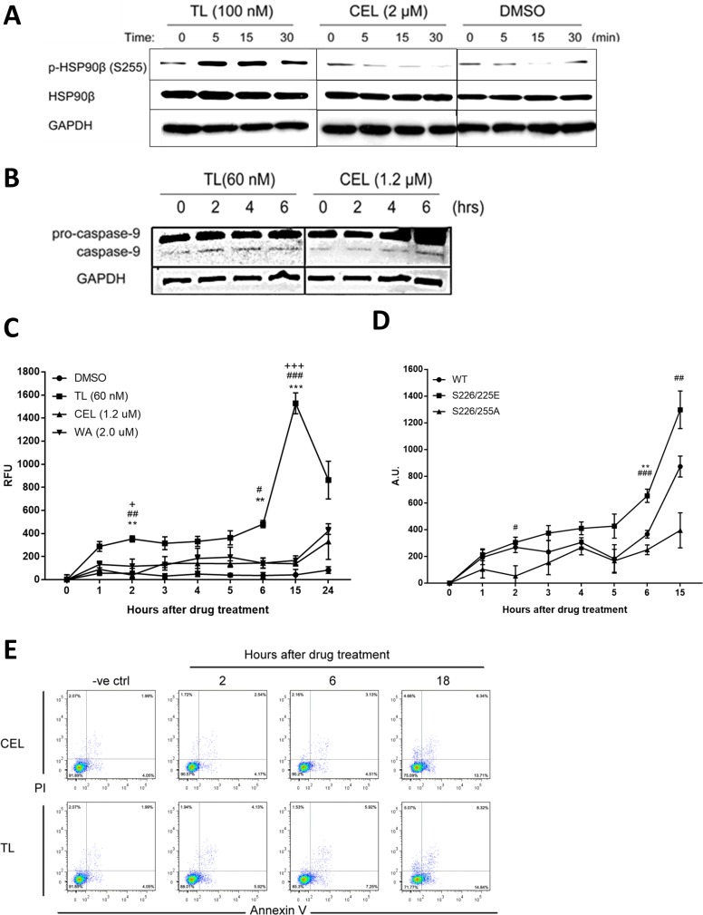
Figure 5. Phosphorylation of HSP90β is induced by TL in early phase.
(A) TL induces site-specific phosphorylation of HSP90β within 5 minutes of drug treatment. Western blot analysis of phosphorylation of HSP90β in HeLa cells treated with TL and CEL for 0, 5, 15 and 30 min. Representative image from three independent experiment was shown. (B) Western blot analysis of caspase-9 in TL/CEL-treated HeLa cells for 0, 2, 4 and 6 hrs. Representative image from three independent experiment was shown. (C) TL activates caspase-3 activity in the early phase. Caspase-3 activity was detected for HeLa cells upon TL, CEL and WA treatment over a time course from 0 to 24 hrs. At indicated time-points, cell lysate was prepared and incubated with assay buffer for at 37°C for 1 hr. Fluorescence was detected with an excitation at 405 nM and emission at 465 nM. **P < 0.01; ***P < 0.001 DMSO versus TL. ##P < 0.01; ###P < 0.001 TL versus CEL. +P < 0.05; +++P < 0.001 TL versus WA. Data are presented as mean ± SD (n = 3). (D) Hypophosphorylation of HSP90β prevents TL mediated early activation of caspase-3 in HeLa cells. Caspase-3 activity assay was performed in HeLa cells transfected with different phosphorylation mutants of HSP90β. Data are presented as mean ± SD (n = 3). **P < 0.01 WT versus S226/225E. #P < 0.05; ##P < 0.01; ###P < 0.001 S226/225E versus S226/225A. (E) TL induces cell apoptosis in a shorter time-frame when compared to those upon CEL treatment. Annexin V/PI analysis of apoptotic HeLa cells upon TL and CEL for 2, 6 and 18 hrs.

