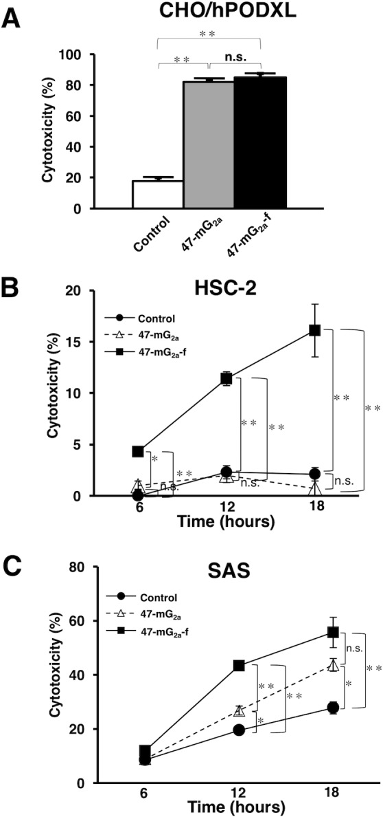
Figure 9. Complement-dependent cytotoxicity (CDC) and antibody-dependent cellular cytotoxicity (ADCC) of anti-PODXL mAbs against PODXL-expressing cell lines.

(A) CDC was evaluated using the 51Cr release assay. Target cells were incubated with [51Cr]sodium chromate (0.1 μCi) for 1 h at 37°C. The 51Cr-labeled cells were incubated with a baby rabbit complement at a dilution of 1:4 in the presence of 47-mG2a, 47-mG2a-f, or control mouse IgG (10 μg/mL) for 6 h in 96-well plates. 51Cr release was measured in the supernatant. (B, C) ADCC was determined using the 51Cr release assay. Target cells such as HSC-2 (B) and SAS (C) were incubated with [51Cr]sodium chromate. After washing, 51Cr-labeled target cells were placed in 96-well plates in triplicate. Effector cells and 47-mG2a, 47-mG2a-f, or control mouse IgG were added to the plates. After incubation (6, 12, and 18 hrs), 51Cr release was measured in the supernatant. The values are means ± SEM. *P < 0.05, **P < 0.01 t-test.
