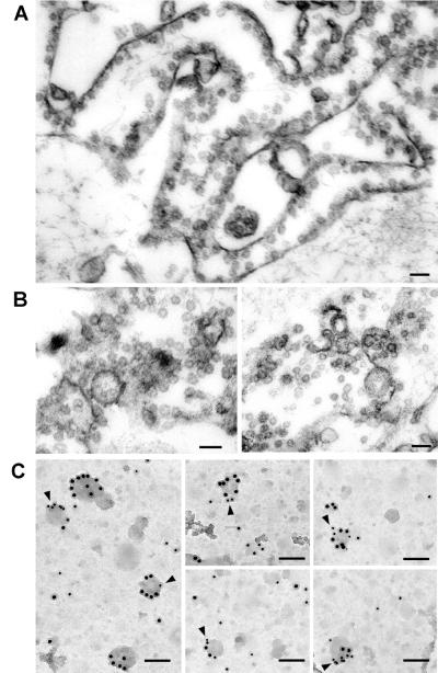
Figure 2.
Electron microscopy of plasma membrane- and caveolae-enriched fractions obtained after caveolae purification from plasma membrane lawns of 3T3-L1 adipocytes. (A) Section of the plasma membrane-enriched fraction harvested from the plates after pulling up caveolae, which shows many caveolae still attached to PM. (Scale bar = 100 nm.) (B) Section of the caveolae-enriched fraction showing many vesicles of homogenous sizes isolated or in grapes. (Scale bar = 100 nm.) (C) Replicas of light caveolin-enriched fractions obtained after centrifugation of the caveolae-enriched fraction overlaid with a sucrose gradient. Membrane fractions were immunolabeled for GLUT4 (monoclonal antibody 1F8 and secondary 10-nm gold-labeled antibody) and caveolin-1 (polyclonal antibody and 15-nm gold-labeled secondary antibody). Arrowheads point to vesicles positive for GLUT4 and caveolin-1. (Scale bar = 85 nm.)

