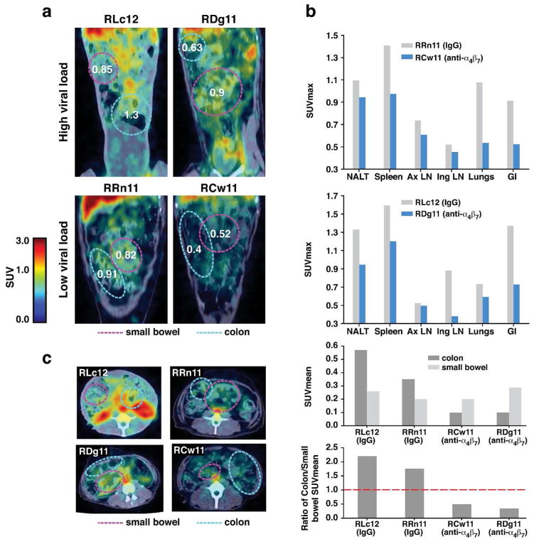
Figure 3. Tissue specific localization of SIV gp120 during early-chronic infection by immuno-PET/CT image analysis.
PET/CT images of the small bowel and colon of 2 macaques (RLC12 and RRN11) that received IgG and 2 macaques that received α4β7 mAb (RDG11 and RCW11) on day −3 and every 3 weeks thereafter until autopsy (weeks 16–17), with 64Cu/DOTA labeled, PEG conjugated SIV gp120 mAb clone 7D3. (a) The immuno-PET/CT frontal images of the 4 macaques, with the small bowel and colon highlighted. (b) SUVmax values for nasal lymphoid tissues (NALT), spleen, axillary lymph-nodes (Ax LN), inguinal lymph-nodes (Ing LN), lungs and the gastro-intestinal tissues (GI) on RRN11 and RCW11 (left bar graph, low viral load) and RLC12 and RDG11 (right bar graph, high viral load). (c) Representative cross-sectional immuno-PET/CT images of SIV for the same 4 macaques is shown (top panel), along with the calculated SUVMean values for the colon and small bowel of the α4β7 mAb (RDG11 and RCW11) and IgG treated (RLC12 and RRN11) animals (middle panel). The ratios of the SUVMean values for the colon versus the small bowel for each of the 4 animals are illustrated (bottom panel). The broken line indicates a ratio of 1.

