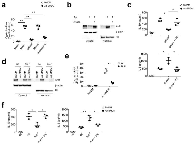
Figure 3. Apoptotic cells activate AhR by a TLR9-dependent mechanism.
(a) Apoptotic cells were treated with DNAse (50 μg/ml) or RNAse (50 μg/ml) prior to culture with BMDM +/− 1μM chloroquine. After 8h Cyp1a1 mRNA was measured by sqPCR. (b) Immunoblot analysis of nuclear and cytoplasmic extracts 2h after Ap-BMDMco-culture +/− DNAse treated apoptotic cells. (c) BMDM were co-cultured with apoptotic cells for 8h (Ap-BMDM) with the indicated conditions alone or in combination with ITE at 1 μM. Culture supernatants were then measured for IL-6 and IL-10 by ELISA. (d) BMDM and Ap-BMDM of the indicated genotype were analyzed by immunoblot analysis of nuclear and cytoplasmic extracts 2h after co-culture as described in b. (e) Cyp1a1 mRNA was measured by sqPCR in BMDM and Ap-BMDM cultures of the indicated genotype. (f) Culture supernatants were measured for the indicated cytokines by ELISA in BMDM and Ap-BMDM after 8h co-culture +/− 1 μM ITE. For all graphs data n=3 biologically independent samples per group and bars in a, c, e, and f are the mean +/− standard deviation. *=P≤ 0.05, **=Pl≤ 0.01, ns= not significant as determined by two-sided Student’s t-test. All experiments were repeated three times with similar results.

