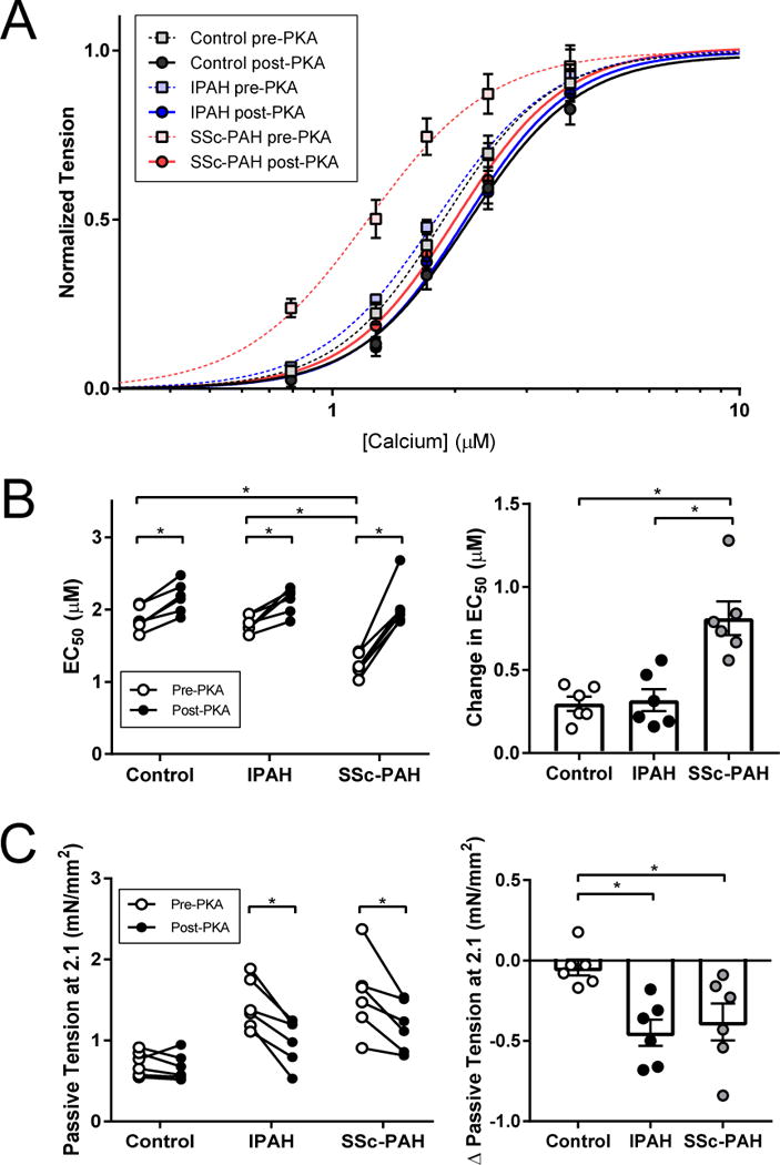
Figure 3.

Effect of PKA treatment on myofilament function and passive myocyte stiffness
(A) Myofilaments from a subset of control, IPAH, and SSc-PAH subjects (3 subjects/group) were directly incubated with protein kinase A (PKA). (B) Baseline calcium sensitivity (EC50) in all three groups mirrored trends of the larger cohort. With PKA, there was a slight increase in EC50 in control and IPAH. On the other hand, PKA completely markedly increased EC50 in the SSc-PAH group to control levels. Change in EC50 was significantly greater in SSc-PAH than in both control and IPAH (Disease × group interaction P=0.002). (C) PKA treatment led to a decrease in passive tension, at a sarcomere length (SL) of 2.1 μm, in both IPAH and SSc-PAH, but no change in controls. There was no significant difference in PKA-mediated passive tension change between IPAH and SSc-PAH. * P<0.05; post-hoc P-value applied where applicable.
