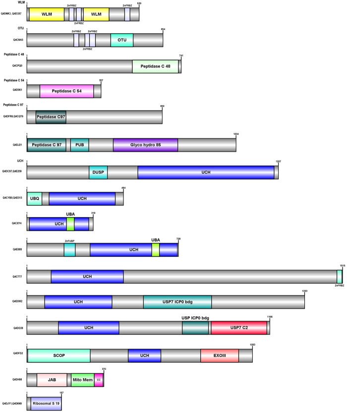
Figure 5.
The Schematic Domain architectures of the Deubiquitinating enzymes (DUBs) proteins in T. cruzi are depicted. The full protein is colored in grey and other domain types are colored in different colors separately. In some of the domain architecture, two domains overlap, so they are represented by two names. The number at top depicts the length of each protein. Domain architecture of atypical UPP proteins based on the presence of unique appended domains or their extensions tethered at N- or C- terminus. Uniprot ids corresponding to each domain diagram is listed at the right. Abbreviations used: SP-signal Peptide, CC-coiled coil.

