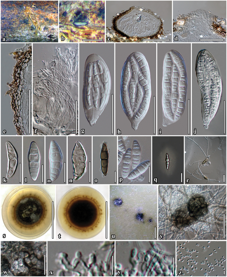
Figure 2.
Neoaquastroma bauhiniae (MFLU 17-0002, holotype) a Appearance of ascomata on host surface b Close up of ascoma c Section of ascoma d Ostiolar canal e Section of partial peridium layer f Pseudoparaphyses g–j Development state of asci j Asci produced in culture k–p Development state of ascospores; (n, o Senescent spores m, p ascospores in 5% of KOH reagent); q Ascospores stained with India ink, sheath surrounding the entire ascospore r Germinated ascospore s, t Culture character on MEA u Conidiomata forming on agar on rice straw media after 8 weeks v Immature conidiomata w Conidiomatal wall x, y Conidiogenous cells and developing conidia z Conidia j, m Asci and ascospore in culture (on rice straw). Scale bars: 500 µm (b); 100 µm (c, v); 50 µm (d–j); 20 µm (k–r, w); 5 µm (x–z).

