Figure 1.
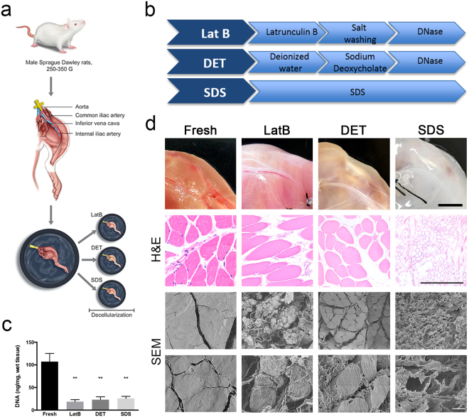
Decellularisation protocols. (a) Diagram showing the process used to prepare rat hind limbs for decellularisation. The femoral artery is cannulated and the hind limb dissected; decellularised reagents where delivered through the vessel tree by using a pump directly connected to the cannula. Three different decellularisation methods were performed, and called LatB, DET and SDS. (b) Diagram showing steps used to perform LatB, DET and SDS decellularisation. (c) Quantification of DNA content in freshly isolated rat skeletal muscle and LatB-, DET- and SDS-decellularised muscles. Data are shown as mean ± s.e.m of three independent replicates; **P < 0.01, one-way ANOVA and Tukey’s multiple comparison test; no significant differences were observed among the decellularised samples; n = 6–10, each group. (d) Macroscopic and microscopic (H&E and scanning electron microscopy, SEM) evaluation of fresh rat muscles and LatB-, DET- and SDS-decellularised samples. For H&E analysis, cross-sections were analysed. Scale bars, macroscopic: 1 cm; H&E: 100 µm; SEM: 10 µm.

