Figure 7.
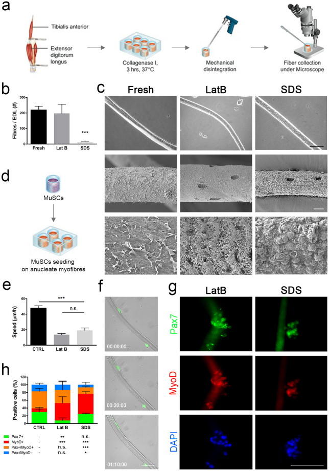
Isolation and seeding of anucleate myofibres. (a) Single myofibres were isolated from fresh or decellularised rat EDL digested in collagenase I and collected under the microscope. (b) Yield of fibres per EDL, ***p < 0.001 vs Fresh, t-test. (c) Scanning electron microscopy of single fresh and anucleate myofibres from LatB- and SDS-decellularised EDL. Scale bar: first row 100 µm, second row 10 µm, third row 1 µm. (d) Murine MuSCs were seeded on single myofibres and fresh EDL fibres were cultured to activate resident MuSCs. (e) Cellular migration speed along single fibres measured 24 hours post-seeding; ***p < 0.001, ANOVA. (f) Representative images of GFP+ MuSCs migrating on LatB-scaffold derived anucleate fibres, scale bar: 10 µm. (g) Immunofluorescence for MyoD and Pax7 of wild-type MuSCs seeded on LatB and SDS anucleate myofibres after 72 hours of culture, scale bar: 100 µm. (h) Subpopulations of cells according to positivity for Pax7 and MyoD after 72 hours of culture with statistical analysis showing significance in respect to control (CTRL). *p < 0.05; **p < 0.01; ***p < 0.001 vs CTRL, t-test.

