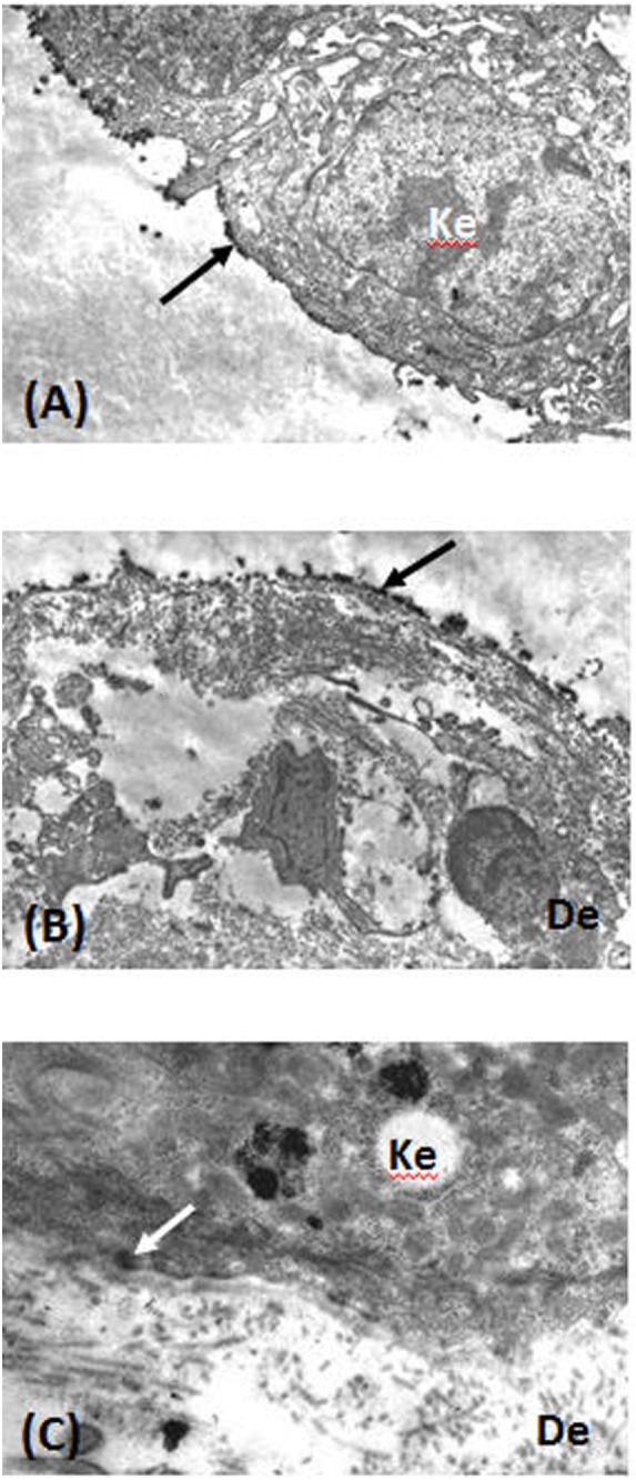
Figure 4.

Direct immunoelectron microscopy of tissue sections from patients 10 (A,B) and 8 (C) was performed as previously described (37). (A) Immune deposits (arrow) on the lamina lucida (LL) cleavage roof. (B) Immune deposits (arrow) in the lower LL and lamina densa at the cleavage floor. (C) Immune deposits (arrow) in the upper LL, close to hemidesmosomes. Abbreviations: Ke, keratinocyte; De, dermis.
