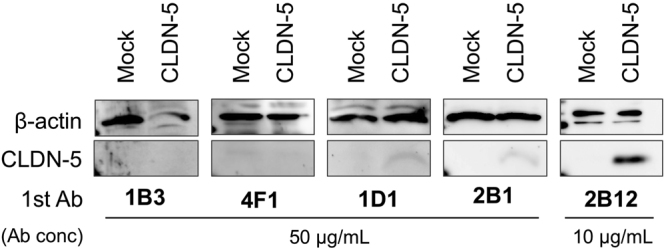
Figure 5.

Binding of anti-CLDN-5 antibody to denatured human CLDN-5. Denatured CLDN-5 was detected by western blotting using anti-CLDN-5 ECR mAbs as primary antibody. Lysates of mock or human CLDN-5–expressing cells were subjected to SDS-PAGE. Blotted membranes were incubated with 10 µg/mL 2B12 or 50 µg/mL the other anti-CLDN-5 ECR mAbs. Regions of CLDN-5 band and β-actin band as a loading control were cropped from original blotting images. Full-length blotting images are shown in Supplementary Fig. S2.
