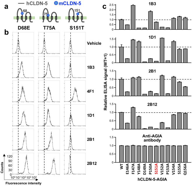
Figure 6.
Epitope mapping of anti-CLDN-5 ECR mAbs. Binding reactivity of anti-CLDN-5 ECR mAbs against HT-1080 cells expressing human/mouse CLDN-5 chimera mutants was examined. (a) Schematic illustration of the human/mouse CLDN-5 mutants. Single amino acid substitution (D68E, T75A, and S151T) was applied to human CLDN-5. The positions of substitution are indicated by gray dots. (b) Flow cytometry. Chimeric mutant expressing cells were treated with vehicle (PBS) or 5 µg/mL of anti-CLDN-5 ECR mAbs. After fluorescein-labeled secondary antibody treatment, fluorescently labeled cells were detected by flow cytometry. (c) Alanine scanning. Alanine mutants were constructed using hCLDN-5-AGIA as template. One of the amino acids from E146 to Q156 was substituted by alanine. Mutants were synthesized on liposome by the wheat cell-free system. Cell-free synthesized proteoliposomes were immobilized on a micro titer plate, and binding between membrane protein and anti-CLDN-5 ECR mAb or anti-AGIA antibody was detected by ELISA.

