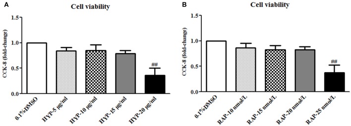
Figure 10.
The cell viability in the cultured mesangial cells. (A) The MCs were exposed to 0.1% DMSO and HYP at 5, 10, 15, and 20 μg/ml for 72 h; (B) The MCs were exposed to 0.1% DMSO and RAP at 10, 15, 20, and 25 nmol/L for 72 h. The data are expressed as mean ± S.E. ##P < 0.01 vs. the treatment of HYP at 15 μg/ml or RAP at 20 nmol/L.

