
Fig. 10.
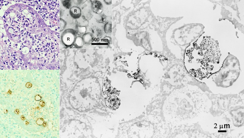
Chlamydial epididymitis immunostained in paraffin sections and processed for EM observation (left top: HE, left bottom and right: immunostaining for chlamydial antigen). Labeled signals are clustered in the dotted inclusion bodies formed in the cytoplasm of epididymal ductal cells. At the EM level, the cell wall of the pathogens shows positivity. At high magnification, large-sized proliferative form (reticulate body: R) and smaller infective form (elementary body: E) are recognized (inset). Bars = 2 μm (inset: 500 nm).