
Fig. 11.
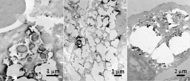
Rods ultrastructurally visualized in the cytoplasm of macrophages in xanthogranulomatous cholecystitis (left), gastric xanthoma (center) and nodal non-tuberculous mycobacteriosis in AIDS (right). Immunostaining using commercial antisera against Escherichia coli (left), Helicobacter pylori (center) and BCG (right) was performed in paraffin sections, respectively. Of note is that the bacilli are packed in the cytoplasm of so-called “striated histiocyte” in the latter lesion. Bars = 1 μm (left and center) and 2 μm (right).