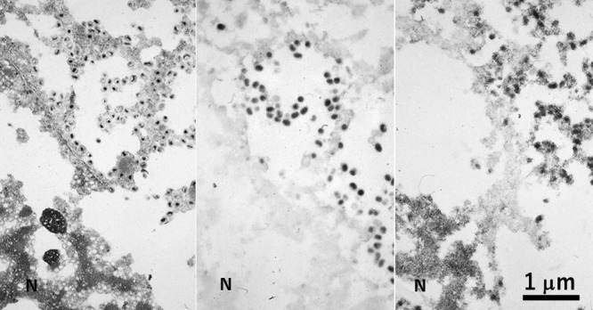
Fig. 12.

A CMV-infected pneumocyte in autopsied and paraffin-embedded lung tissue consecutively observed by conventional EM (left), immunostaining (center) and ISH (right). In spite of poor morphologic preservation, enveloped cored viral particles are clearly seen in the cytoplasm, while the nuclei are packed with virions without envelope. Immunoelectron microscopy using an antiserum against envelope protein discloses enveloped particles in the cytoplasm, while ISH detects the viral genome in both the cytoplasmic viral particles and envelope-less virions clustered in the nuclear matrix. N = nucleus. Bar = 1 μm.
