
Fig. 13.
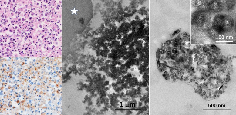
Ultrastructural detection of viral particles in the lymph node of SFTS in formalin-fixed, paraffin-embedded sections (left top: HE, left bottom and center: immunostaining using a monoclonal antibody clone 1C3, right: ISH by the AT-tailing method using biotinylated antisense cocktail probes). Necrotizing lymphadenitis-like features are discerned in the enlarged node. Viruses are distributed in the cytoplasm of hemophagocytic macrophages. At the ultrastructural level, the labeled viral particles are round in shape and around 100 nm in size. Asterisk indicates a phagocytized erythrocyte. TACASTM slides allowed both the prevention of slide detachment during heating pretreatment and transfer of the stained paraffin section to the Epon block. Bars = 1 μm (center), 500 nm (right) and 100 nm (inset).