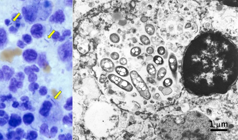
Fig. 2.

Legionnaire’s pneumonia fixed in unbuffered formalin (left: Giemsa stain of touch smear of the autopsied lung lesion, right: EM). In the cytoplasm of macrophages infiltrating in lobar pneumonia, rods are observed (arrows). Ultrastructurally, rod-shaped bacteria corresponding to Legionella pneumophila are phagocytized in lysosomes. Nuclear chromatin pattern is well preserved. Bar = 1 μm.
