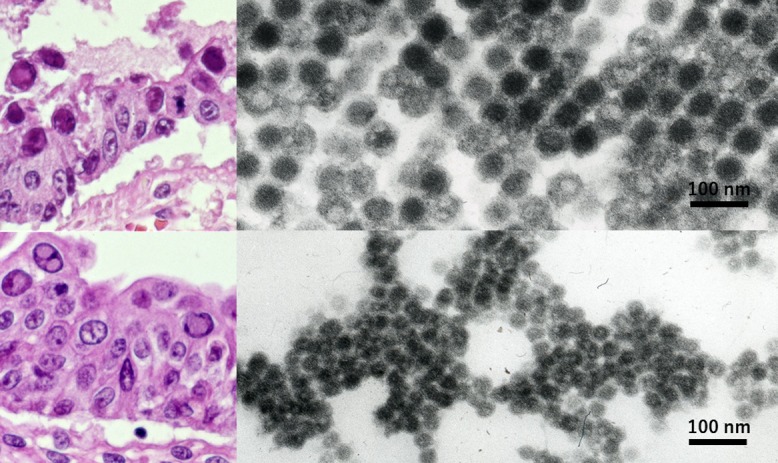
Fig. 5.

Opportunistic dual infection of adenovirus and BK virus ultrastructurally identified in formalin-fixed, paraffin-embedded sections of autopsy material (left: HE, right: EM, top: small bowel mucosa, bottom: urinary bladder mucosa). Nuclear inclusion bodies of both smudge and cored types are indistinguishable at the light microscopic level. EM evaluation using paraffin sections contributed to identifying two different DNA viruses in the nuclei. Adenovirus larger than BK virus is hexagonal on cut surface and regularly arranged. Some adenoviral particles are vacant. Bars = 100 nm.
