Fig. 8.

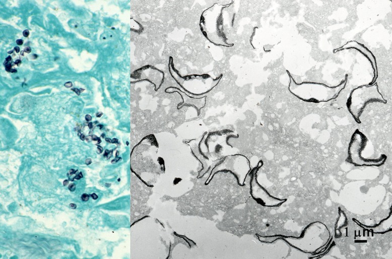
Grocott stain in pneumocystosis in paraffin sections of lung biopsy at the light and electron microscopic levels. The cyst wall of Pneumocystis jirovecii is densely silver-impregnated. Alveolar space is filled with the black-signaled, crescent-shaped pathogen. Dot-like signals as focal thickening of the cell wall are observed at both light and electron microscopic levels. Bar = 1 μm.
