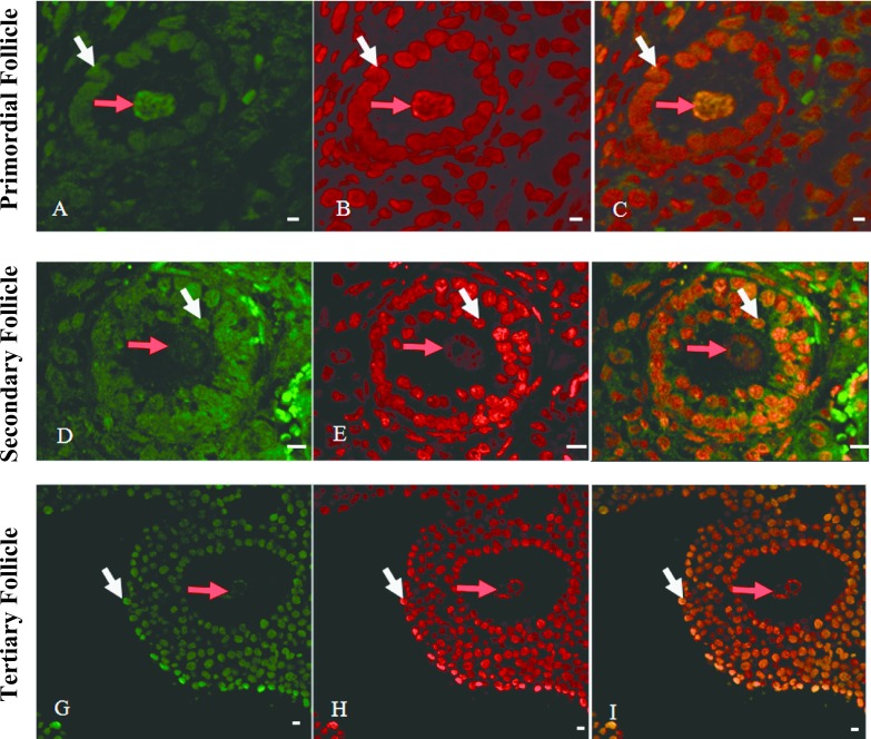
Fig. 4.
Simultaneous localization of non-methylated and methylated GATCG sequences by HELMET in a paraffin-embedded section of mouse ovary. The paraffin-embedded section was blocked with a dideoxynucleotide mixture by TdT and then the Sau3A I cutting sites were labeled with biotin-16-dUTP. After dideoxynucleotide blockade, Mbo I cutting sites were labeled with digoxigenin-11-dUTP and then both haptens were visualized with FITC-anti-biotin (A, D and G) and rhodamine-anti-digoxigenin (B, E and H), respectively. Merged images are given in C, F and I. Red arrow pointed the nuclei of oocytes and white arrow pointed granulosa cells. And non-methylated GATCG was localized in the nuclei of granulosa cells (white arrows) and its staining became to be more intense from primary to secondary follicles. Bar = 20 μm.

