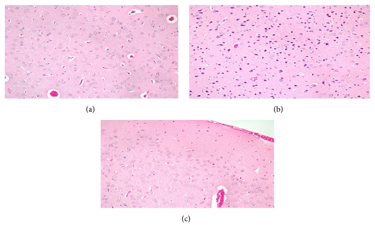
Figure 3.
(a) Histopathological image of section of the brain of a control rat. The image shows normal cells in the brain. (b) Histopathological image of section of the brain of rat treated with AlCl3. It shows numerous small dark cells with no nucleus which are apoptotic cells. (c) Histopathological image of section of the brain of AlCl3 exposed rats pretreated with quercetin and α-lipoic acid treated rat. It shows normal cells with fewer apoptotic cells.

