Figure 6.
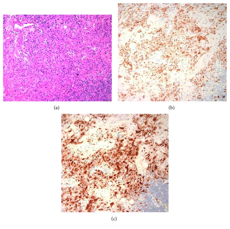
Non-Langerhans cell histiocytosis: (a) low-power (20x) image shows a histiocytic proliferation which is diffusely positive for CD68 and resembles that of Langerhans cell histiocytosis but with a comparatively low amount of eosinophils and negative immunostaining for CD1a. (b) CD68+. (c) Factor XIII+.

