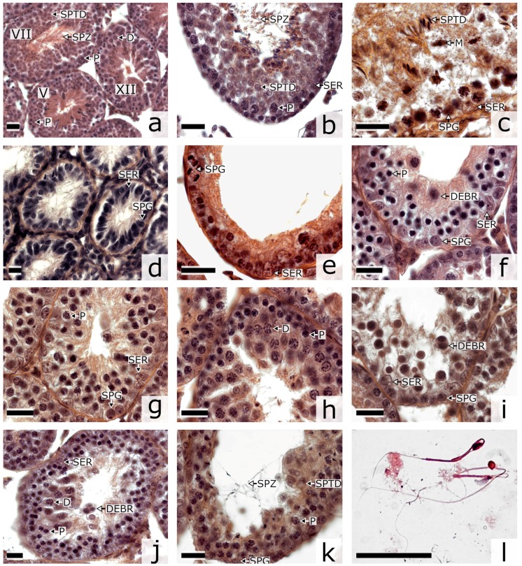
Figure 1.
Histological sections of testes of Phodopus campbelli (a–c) and F1 type A (d,e) and type B (f–l) hybrids stained by hematoxylin-eosin. D, diplotene-diakinesis; DEBR, cellular debris; M, metaphase I and II; P, pachytene; SER, Sertoli cell; SPG, spermatogonium; SPTD, spermatid; SPZ, spermatozoid; E, early spermatocyte I at leptotene-zygotene. (a) tubules at different stages of the seminiferous epithelium cycle (V, VII and XII) showing an undisturbed progression of the spermatogenic wave; (b) stage VIII tubule containing Sertoli cells, spermatocytes I at pachytene and spermatids, its lumen containing mature spermatozoa; (c) stage XIII-XIV tubule containing spermatocytes at metaphase I and II and spermatids at the upper layer and Sertoli cells and spermatogonia at the lower layer; (d) aberrant seminiferous tubules without spermatogenic epithelium, (e) wall of an empty tubule with dividing spermatogonia and Sertoli cells only; (f) tubule with an excess of early spermatocytes I at leptotene and zygotene at the lower layer, cellular debris at the upper layer; (g) tubule with an excess of spermatocytes I at pachytene; (h) tubule wall with an excess of spermatocytes I at diplotene-diakinesis; (i) tubule containing degenerating pachytene spermatocytes and cellular debris; (j) tubule containing degenerating diplotene-diakinesis spermatocytes and cellular debris; (k) tubule with abnormal spermatids in the wall and abnormal spermatozoa in the lumen; (l) abnormal spermatozoa in the epididymal smear. Bar: 20 µm.

