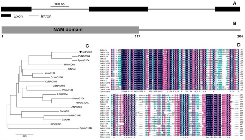
Figure 1.
Gene structure and protein sequence analysis of MdNAC1. (A) Exons/introns of MdNAC1 are displayed. (B) NAM domain detected in the N-terminus region of MdNAC1. (C) Phylogenetic analysis of MdNAC1 (marked by solid-black circle) and homologous proteins from other species. (D) Alignment of MdNAC1 and its homologous proteins. These different colors show the similarity degrees of the amino acids. Homologous protein sequences of MdNAC1 from 18 other species were used for phylogenetic analysis and alignment.

