Figure 3.
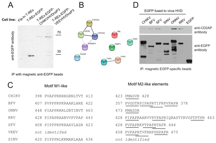
Binding of CD2AP by nsP3 HVDs of different alphaviruses. (A) Western blot analysis of immunoprecipitated samples obtained from Flp-In T-Rex, T-Rex-EGFP, T-Rex-EGFP-CHIKVnsP3HVD and T-Rex-EGFP-VEEVnsP3HVD cells with anti-EGFP antibody; (B) Network analysis of cellular proteins captured by VEEV nsP3 HVD performed as described for Figure 1C; (C) M1 and M2 like elements identified via multiple sequence alignment in nsP3 HVD of different Old and New World alphaviruses. Underline indicates CHIKV CD2AP-binding M2 motif identified in this study or a motif similar to it; (D) Flp-In T-REx cells were transfected with plasmids expressing EGFP fused to HVD from the indicated Old World alphaviruses; 24 h p.t. cells were lysed and EGFP-specific magnetic beads were used to pull down proteins; obtained samples were probed for presence of CD2AP using antibody against CD2AP. Blot developed using anti-EGFP antibody is shown as recombinant protein expression/capture control.

